
Comparative overview and why it matters
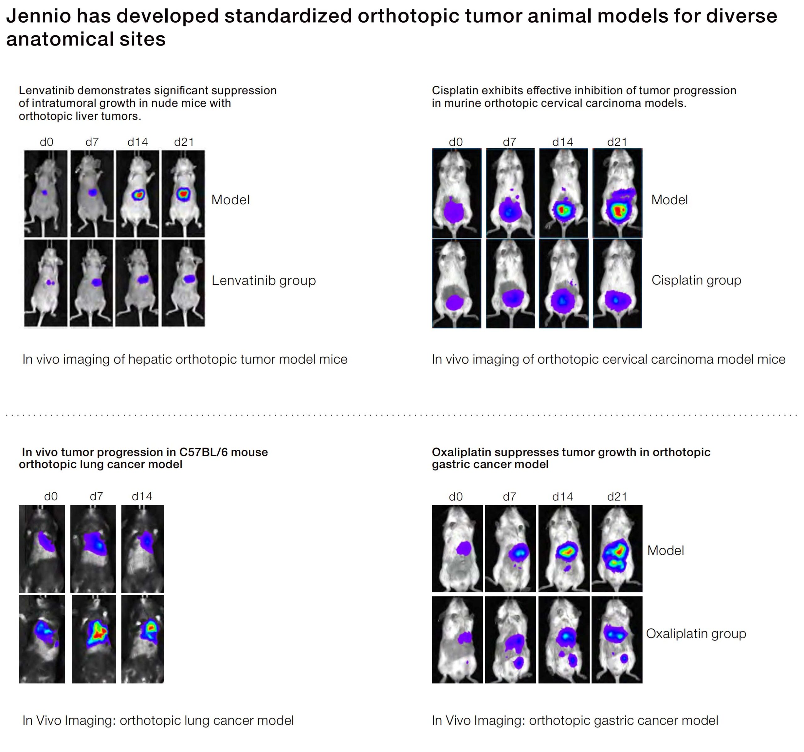
Direct comparisons clarify what a model actually predicts. Bench scientists, pharmacologists and study designers repeatedly face a trade-off between throughput and biological fidelity. An orthotopic tumor model places tumor cells in their organ of origin, increasing relevance for invasion and metastatic phenotypes, whereas alternative loci speed screening but reduce physiological context. This piece takes a comparative-insight lens to orthotopic vs subcutaneous tumor model choices, focusing on actionable design, measurable endpoints and operational constraints.
Design and biological fidelity
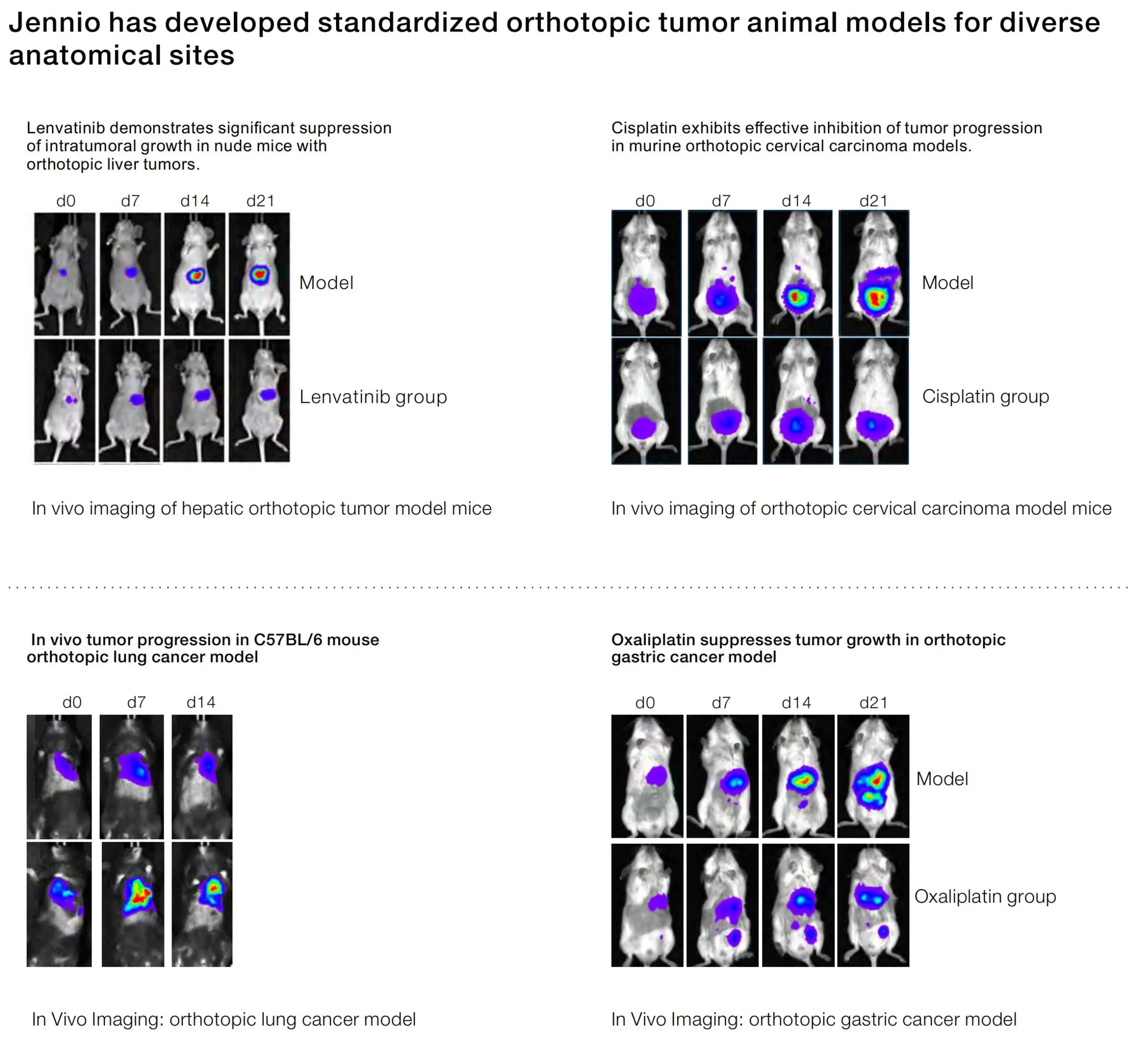
Orthotopic implantation recreates native stromal interactions, permitting evaluation of the tumor microenvironment and realistic metastasis routes. Subcutaneous xenografts simplify measurement — palpable volume changes and consistent growth curves — but miss organ-specific vasculature, extracellular matrix composition and immune cell trafficking. Where immune context is pivotal, syngeneic orthotopic models or humanized xenografts give superior translational signal; for mechanism-of-action screens, subcutaneous models remain efficient. Use of bioluminescence imaging or MRI aligns with orthotopic setups when internal tumor burden is the primary endpoint.
Operational production teardown: resources, throughput, and integration
Operational planning must map to the biological objective. In a production teardown, quantify these inputs: surgical labor per implantation, perioperative mortality rate target (<2–5% for experienced teams), imaging cadence and assay turnaround. Embed orthotopic tumor model and orthotopic vs subcutaneous tumor model directly into study SOPs so procurement, anesthesia protocols and endpoint definitions are synchronized. Tissue processing (single-cell prep, IHC panels) requires different sampling windows for orthotopic tumors due to heterogeneous necrosis patterns. Plan cage-level randomization and blinding early — these reduce bias without adding assay time.
Readouts, analytics, and validation — matching assay to hypothesis
Choose readouts that align with mechanism and regulatory expectations. Pharmacodynamic biomarkers collected from orthotopic lesions often reflect tumor-stroma pharmacokinetics more accurately. For safety and dose-finding, subcutaneous volume metrics support rapid dose–response modeling. Validate translational assumptions against an external anchor: the National Cancer Institute’s patient-derived xenograft (PDX) repository has been used by multiple centers to correlate preclinical responses with clinical outcomes, demonstrating that orthotopic placement frequently increases predictive value for metastatic behavior. Incorporate multiplexed histology or RNA expression panels when you require pathway-level confirmation.
Common pitfalls, alternatives, and corrective actions
Underpowered cohort sizes and inconsistent implantation technique are the two most common failure modes. Avoid mixing implantation routes within a single study — it confounds growth kinetics and immune readouts. If surgical expertise is limited, consider organoid-based orthotopic grafts or refined subcutaneous controls as staging models. For labs transitioning from subcutaneous screens to orthotopic validation, run parallel pilot arms to calibrate imaging thresholds and humane endpoints — this reduces downstream variance. Also note: variability in tumor take-rate often necessitates oversampling by 10–30% based on historical throughput.
Advisory: three golden rules for model selection
1) Align the model to the decision gate: use orthotopic models for invasion, metastasis or microenvironment-dependent mechanism validation; use subcutaneous models for initial efficacy and high-throughput dose finding. 2) Quantify operational burden: require explicit metrics — implantation labor hours per cohort, imaging frequency, and expected sample processing time — before committing to study scale. 3) Lock assay endpoints to translational anchors: histopathology scoring, circulating tumor DNA trends or established PDX correlations must be pre-specified and statistically powered.
Choosing the right in vivo model reduces downstream risk and sharpens go/no-go decisions; when that decision points toward higher biological fidelity, the value proposition of standardized orthotopic workflows becomes clear. For teams needing integrated orthotopic platforms and validated assay pipelines, Jennio Biotech supplies reproducible implants and support aligned to translational objectives — a practical way to close the experimental loop. —